
Chapter 7: The Respiratory System
Learning Objectives
By the end of this section, you will be able to:
- Describe the passage of air from the outside environment to the lungs
- Describe the function of the respiratory system
Take a breath in and hold it. Wait several seconds and then let it out. Humans, when they are not exerting themselves, breathe approximately 15 times per minute on average. This equates to about 900 breaths an hour or 21,600 breaths per day. With every inhalation, air fills the lungs, and with every exhalation, it rushes back out. That air is doing more than just inflating and deflating the lungs in the chest cavity. The air contains oxygen that crosses the lung tissue, enters the bloodstream, and travels to organs and tissues. There, oxygen is exchanged for carbon dioxide, which is a cellular waste material. Carbon dioxide exits the cells, enters the bloodstream, travels back to the lungs, and is expired out of the body during exhalation.
The Human Respiratory System
Breathing is both a voluntary and an involuntary event. How often a breath is taken and how much air is inhaled or exhaled is regulated by the respiratory center in the brain in response to signals it receives about the carbon dioxide content of the blood. However, it is possible to override this automatic regulation for activities such as speaking, singing and swimming under water.
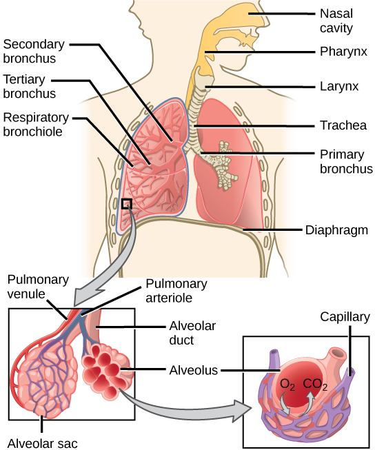
In humans, pulmonary ventilation occurs via inhalation (breathing). During inhalation, air enters the body through the nasal cavity located just inside the nose (Figure 7.1). As air passes through the nasal cavity, the air is warmed to body temperature and humidified. The respiratory tract is coated with mucus to seal the tissues from direct contact with air. Mucus is high in water. As air crosses these surfaces of the mucous membranes, it picks up water. These processes help equilibrate the air to the body conditions, reducing any damage that cold, dry air can cause. Particulate matter that is floating in the air is removed in the nasal passages via mucus and cilia. The processes of warming, humidifying, and removing particles are important protective mechanisms that prevent damage to the trachea and lungs. Air is also chemically sampled by the sense of smell. Thus, inhalation serves several purposes in addition to bringing oxygen into the respiratory system.
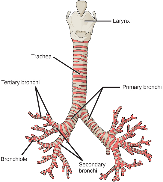
From the nasal cavity, air passes through the pharynx (throat) and the larynx (voice box), as it makes its way to the trachea (Figure 7.2). The main function of the trachea is to funnel the inhaled air to the lungs and the exhaled air back out of the body. The human trachea is a cylinder about 10 to 12 cm long and 2 cm in diameter that sits in front of the esophagus and extends from the larynx into the chest cavity where it divides into the two primary bronchi. It is made of incomplete rings of cartilage and smooth muscle. The cartilage provides strength and support to the trachea to keep the passage open. The trachea is lined with cells that have cilia and secrete mucus. The mucus catches particles that have been inhaled, and the cilia move the particles toward the pharynx. The cartilage provides strength and support to the trachea to keep the passage open. The smooth muscle can contract, decreasing the trachea’s diameter, which causes expired air to rush upwards from the lungs at a great force. The forced exhalation helps expel mucus when we cough. Smooth muscle can contract or relax, depending on stimuli from the external environment or the body’s nervous system.
Lungs: Bronchi and Alveoli
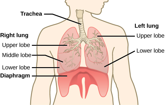
The end of the trachea bifurcates (divides) to the right and left lungs. The lungs are not identical. The right lung is larger and contains three lobes, whereas the smaller left lung contains two lobes (Figure 7.3). The muscular diaphragm, which facilitates breathing, is below the lungs and marks the end of the thoracic cavity.
In the lungs, air is diverted into smaller and smaller passages, or bronchi. Air enters the lungs through the two primary (main) bronchi (singular: bronchus). Each bronchus divides into secondary bronchi, then into tertiary bronchi, which in turn divide, creating smaller and smaller diameter bronchioles as they split and spread through the lung. Like the trachea, the bronchi are made of cartilage and smooth muscle. At the bronchioles, the cartilage is replaced with elastic fibers. Bronchi are innervated by nerves of both the parasympathetic and sympathetic nervous systems that control muscle contraction (parasympathetic) or relaxation (sympathetic) in the bronchi and bronchioles, depending on the nervous system’s cues. In humans, bronchioles with a diameter smaller than 0.5 mm are the respiratory bronchioles. They lack cartilage and therefore rely on inhaled air to support their shape. As the passageways decrease in diameter, the relative amount of smooth muscle increases.
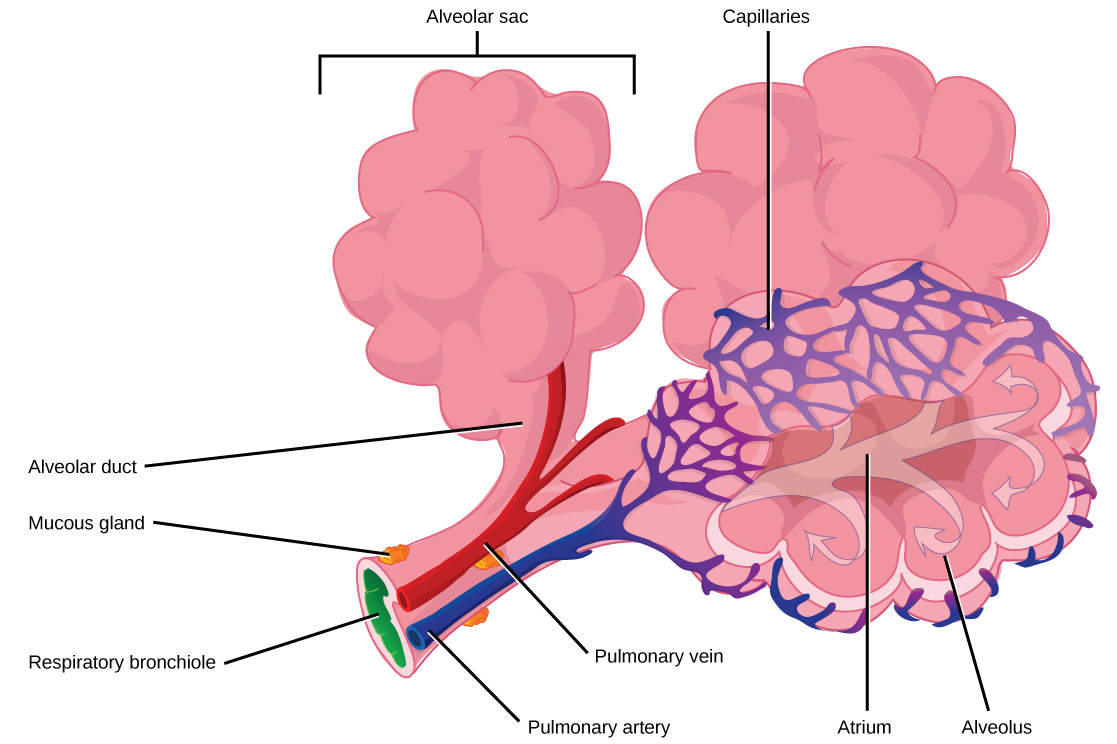
The terminal bronchioles subdivide into microscopic branches called respiratory bronchioles. The respiratory bronchioles subdivide into several alveolar ducts. Numerous alveoli and alveolar sacs surround the alveolar ducts. The alveolar sacs resemble bunches of grapes tethered to the end of the bronchioles (Figure 7.4). Gas exchange occurs only in alveoli. The alveoli are thin-walled and look like tiny bubbles within the sacs. The alveoli are in direct contact with capillaries (very small blood vessels) of the circulatory system. Such intimate contact ensures that oxygen will diffuse (move) from the alveoli into the blood. In addition, carbon dioxide will diffuse from the blood into the alveoli to be exhaled. The anatomical arrangement of capillaries and alveoli emphasizes the structural and functional relationship of the respiratory and circulatory systems.
Because there are so many alveoli (~300 million per lung) within each alveolar sac and so many sacs at the end of each alveolar duct, the lungs have a sponge-like consistency. Estimates for the surface area of alveoli in the lungs vary around 100 m2. This large area is about the area of half a tennis court. This large surface area, combined with the thin-walled nature of the alveolar cells, allows gases to easily diffuse across the cells. The primary function of the respiratory system is to deliver oxygen to the cells of the body’s tissues and remove carbon dioxide, a cell waste product. The main structures of the human respiratory system are the nasal cavity, the trachea, and lungs.
Protective Mechanisms
The air that organisms breathe contains particulate matter such as dust, dirt, viral particles, and bacteria that can damage the lungs or trigger allergic immune responses. The respiratory system contains several protective mechanisms to avoid problems or tissue damage. In the nasal cavity, hairs and mucus trap small particles, viruses, bacteria, dust, and dirt to prevent their entry.
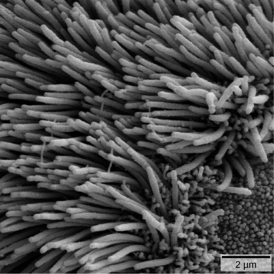
If particulates do make it beyond the nose, or enter through the mouth, the bronchi and bronchioles of the lungs also contain several protective devices. The lungs produce mucus—a sticky substance made of mucin, a complex glycoprotein, as well as salts and water—that traps particulates. The bronchi and bronchioles contain cilia, small hair-like projections that line the walls of the bronchi and bronchioles (Figure 7.5). These cilia beat in unison and move mucus and particles out of the bronchi and bronchioles back up to the throat where it is swallowed and eliminated via the esophagus.
Tar and other substances in cigarette smoke destroy or paralyze the cilia, making the removal of particles more difficult. In addition, smoking causes the lungs to produce more mucus, which the damaged cilia are not able to move. This causes a persistent cough, as the lungs try to rid themselves of particulate matter, and makes smokers more susceptible to respiratory ailments.
Summary
The human respiratory system is designed to facilitate gas exchange. Air is warmed and humidified in the nasal cavity, then travels down the pharynx, through the trachea, and into the lungs. In the lungs, air passes through the branching bronchi, reaching the respiratory bronchioles, which house the first site of gas exchange. The respiratory bronchioles open into the alveolar ducts, alveolar sacs, and alveoli. Because there are so many alveoli and alveolar sacs in the lung, the surface area for gas exchange is very large. Several protective mechanisms are in place to prevent damage or infection. These include the hair and mucus in the nasal cavity that trap dust, dirt, and other particulate matter before they can enter the system. In the lungs, particles are trapped in a mucus layer and transported via cilia up to the esophageal opening at the top of the trachea to be swallowed.
Exercises
- Which of the following statements about the human respiratory system is false?
- When we breathe in, air travels from the pharynx to the trachea.
- The bronchioles branch into bronchi.
- Alveolar ducts connect to alveolar sacs.
- Gas exchange between the lungs and blood takes place in the alveolus.
- The respiratory system ________.
- provides body tissues with oxygen
- provides body tissues with oxygen and carbon dioxide
- establishes how many breaths are taken per minute
- provides the body with carbon dioxide
- Which is the order of airflow during inhalation?
- nasal cavity, trachea, larynx, bronchi, bronchioles, alveoli
- nasal cavity, larynx, trachea, bronchi, bronchioles, alveoli
- nasal cavity, larynx, trachea, bronchioles, bronchi, alveoli
- nasal cavity, trachea, larynx, bronchi, bronchioles, alveoli
- Describe the function of these terms and describe where they are located: main bronchus, trachea, alveoli.
- How does the structure of alveoli maximize gas exchange?
Answers
- B
- A
- B
- The main bronchus is the conduit in the lung that funnels air to the airways where gas exchange occurs. The main bronchus attaches the lungs to the very end of the trachea where it bifurcates. The trachea is the cartilaginous structure that extends from the pharynx to the lungs. It serves to funnel air to the lungs. The alveoli are the site of gas exchange; they are located at the terminal regions of the lung and are attached to the alveolar sacs, which come from the alveolar ducts and respiratory bronchioles terminal bronchi.
- The sac-like structure of the alveoli increases their surface area. In addition, the alveoli are made of thin-walled cells. These features allows gases to easily diffuse across the cells.
Glossary
alveolus: (plural: alveoli) (also, air sacs) the terminal structure of the lung passage where gas exchange occurs
bronchi: (singular: bronchus) smaller branches of cartilaginous tissue that stem off of the trachea; air is funneled through the bronchi to the region where gas exchange occurs in the alveoli
bronchiole: an airway that extends from the main bronchus to the alveolar sac
diaphragm: a skeletal muscle located under lungs that encloses the lungs in the thorax
larynx: the voice box, located within the throat
nasal cavity: an opening of the respiratory system to the outside environment
pharynx: the throat
primary bronchus: (also, main bronchus) a region of the airway within the lung that attaches to the trachea and bifurcates to form the bronchioles
trachea: the cartilaginous tube that transports air from the throat to the lungs
