19 Female Reproductive System
Learning Objectives
- Identify the anatomy of the female reproductive system
- Describe the main functions of the female reproductive system
- Spell the medical terms of the female reproductive system and use correct abbreviations
- Identify the medical specialties associated with the female reproductive system
- Explore common diseases, disorders, and procedures related to the female reproductive system
Female Reproductive System Word Parts
Click on prefixes, combining forms, and suffixes to reveal a list of word parts to memorize for the female reproductive system.
Introduction to the Female Reproductive System
The female reproductive system produces gametes and reproductive hormones. In addition, the female reproductive system supports the developing fetus and delivers it to the outside world. The female reproductive system is located primarily inside the pelvic cavity. The female gonads are called ovaries and the gamete they produce is called an oocyte.

Watch this video:
Media 10.1. Reproductive System, Part 1 – Female Reproductive System: Crash Course A&P #40 [Online video]. Copyright 2015 by CrashCourse.
Female Reproductive System Medical Terms
Anatomy (Structures) of the Female Reproductive System
External Female Genitals
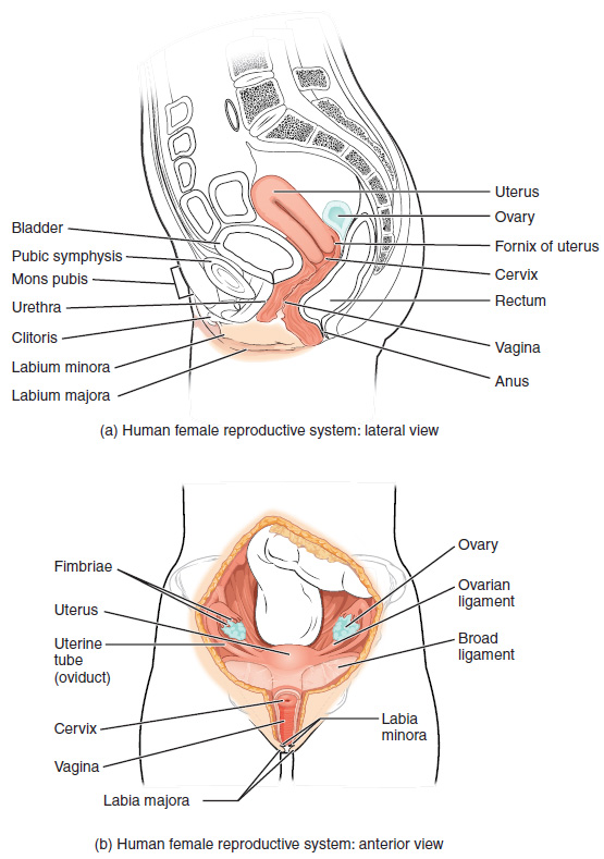
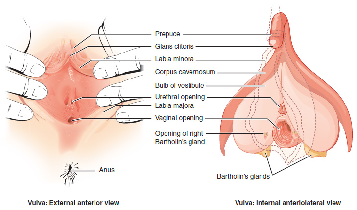
The external female reproductive structures are referred to collectively as the vulva and they include:
- The mons pubis is a pad of fat that is located at the anterior, over the pubic bone. After puberty, it becomes covered in pubic hair.
- The labia majora (labia = “lips”; majora = “larger”) are folds of hair-covered skin that begin just posterior to the mons pubis.
- The labia minora (labia = “lips”; minora = “smaller”) is thinner and more pigmented and extends medially to the labia majora.
- Although they naturally vary in shape and size from woman to woman, the labia minora serve to protect the female urethra and the entrance to the female reproductive tract.
- The superior, anterior portions of the labia minora come together to encircle the clitoris (or glans clitoris), an organ that originates from the same cells as the glans penis and has abundant nerves that make it important in sexual sensation and orgasm. The hymen is a thin membrane that sometimes partially covers the entrance to the vagina.
- The vaginal opening is located between the opening of the urethra and the anus. It is flanked by outlets to the Bartholin’s glands.

Internal Female Reproductive Organs
Vagina
The vagina is a muscular canal (approximately 10 cm long) that is the entrance to the reproductive tract. It also serves as the exit from the uterus during menses and childbirth. The outer walls of the anterior and posterior vagina are columns with ridges. The superior fornix meets the uterine cervix. The cervix is the opening to the uterus.
The walls of the vagina are lined with:
- An outer, fibrous adventitia
- A middle layer of smooth muscle
- An inner mucous membrane with transverse folds called rugae.
Together, the middle and inner layers allow the expansion of the vagina to accommodate intercourse and childbirth. The thin, perforated hymen can partially surround the opening to the vaginal orifice. The Bartholin’s glands and the lesser vestibular glands (located near the clitoris) secrete mucus, which keeps the vestibular area moist.
The vagina has a normal population of microorganisms that help to protect against infection. There is both pathogenic bacteria, and yeast in the vagina. In a healthy woman, the most predominant type of vaginal bacteria is from the genus Lactobacillus, which secretes lactic acid. the lactic acid protects the vagina by maintaining an acidic pH (below 4.5).
Lactic acid, in combination with other vaginal secretions, makes the vagina a self-cleansing organ. However, douching can disrupt the normal balance of healthy microorganisms, and increase a woman’s risk for infections and irritation. It is recommend that women do not douche and that they allow the vagina to maintain its normal healthy population of protective microbial flora.
Ovaries
The ovaries are the female gonads. There are two, one at each entrance to the fallopian tube. They are each about 2 to 3 cm in length, about the size of an almond. The ovaries are located within the pelvic cavity. The ovary itself is attached to the uterus via the ovarian ligament. The ovarian stroma forms the bulk of the adult ovary. Oocytes develop within the outer layer of this stroma, each surrounded by supporting cells. This grouping of an oocyte and its supporting cells is called a follicle.
The Fallopian Tubes
The fallopian tubes are the conduit of the oocyte from the ovary to the uterus. Each of the two fallopian tubes is close to, but not directly connected to, the ovary.
- The isthmus is the narrow medial end of each uterine tube that is connected to the uterus.
- The wide distal infundibulum flares out with slender, finger-like projections called fimbriae.
- The middle region of the tube, called the ampulla, is where fertilization often occurs.
The fallopian tubes have three layers:
- An outer serosa
- A middle smooth muscle layer
- An inner mucosal layer
- In addition to its mucus-secreting cells, the inner mucosa contains ciliated cells that beat in the direction of the uterus, producing a current that will be critical to moving the oocyte .
The Uterus and Cervix
The uterus is the muscular organ that nourishes and supports the growing embryo. Its average size is approximately 5 cm wide by 7 cm long and it has three sections.
- The portion of the uterus superior to the opening of the uterine tubes is called the fundus.
- The middle section of the uterus is called the body of uterus (or corpus).
- The cervix is the narrow inferior portion of the uterus that projects into the vagina.
- The cervix produces mucus secretions that become thin and stringy under the influence of high systemic plasma estrogen concentrations, and these secretions can facilitate sperm movement through the reproductive tract.
The wall of the uterus is made up of three layers:
- Perimetrium: the most superficial layer and serous membrane.
- Myometrium: a thick layer of smooth muscle responsible for uterine contractions.
- Endometrium: the innermost layer containing a connective tissue lining covered by epithelial tissue that lines the lumen. It provides the site of implantation for a fertilized egg, and sheds during menstruation if no egg is fertilized.
Concept Check
- Write or draw out the components of the pathway that an oocyte takes from beginning to end.
- Why do you think the fallopian tubes are not connected to the ovaries?
Physiology (Function) of the Female Reproductive System-Ovulation
Following ovulation, the Fallopian tube receives the oocyte. Oocytes lack flagella, and therefore cannot move on their own.
- High concentrations of estrogen that occur around the time of ovulation induce contractions of the smooth muscle along the length of the Fallopian tube.
- These contractions occur every 4 to 8 seconds, causing the oocyte to flow towards the uterus, through the coordinated beating of the cilia that line the outside and lumen of the length of the Fallopian tube which pulls the oocyte into the interior of the tube.
- Once inside, the muscular contractions and beating cilia move the oocyte slowly toward the uterus.
- When fertilization does occur, sperm typically meet the egg while it is still moving through the ampulla.
Watch this video on ovulation from MedLine Plus to observe ovulation and its initiation in response to the release of FSH and LH from the pituitary gland.[1]
The Menstrual Cycle
The three phases of the menstrual cycle are:
- The menses phase of the menstrual cycle is the phase during which reproductive hormone levels are low, the woman menstruates, and the lining is shed. The menses phase lasts between 2 – 7 days with an average of 5 days.
- The proliferative phase is when menstrual flow ceases and the endometrium begins to proliferate . During this phase reproductive hormones are working in homeostasis to trigger ovulation on approximately day 14 of a typical 28-day menstrual cycle. Ovulation marks the end of the proliferative phase.
- The secretory phase the endometrial lining prepares for implantation of a fertilized egg. If no pregnancy occurs within approximately 10- 12 days the endometrium will grow thinner and shed starting the first day of the next cycle.
Anatomy Labeling Activity
Female Reproductive System Terms not Easily Broken into Word Parts
Female Reproductive System Medical Abbreviations
Diseases and Disorders of the Female Reproductive System
Cancer
Breast Cancer
Breast cancer starts in the cells that line the ducts or the lobule of the breast. Some warning signs include a new lump in the breast or axilla, thickening or swelling, irritation or dimpling of the breast skin, redness or flaky skin, pain, discharge, all in the breast or nipple area, and change in breast size. Risk factors include family history, obesity, hormonal treatment and changes in breast cancer-related genes (BRCA1 or BRCA2) (Centers for Disease Control and Prevention, n.d.; Cancer Care Ontario, n.d.).
Treatment options include chemotherapy, radiation and surgical interventions such as mastectomy, biopsy, incision and drainage and mammoplasty (Centers for Disease Control and Prevention, n.d.; Cancer Care Ontario, n.d.). To learn more about breast cancer, view the Cancer Care Ontario: Breast Cancer web page.
Cervical Cancer
Cervical cancer is typically slow-growing cancer and is highly curable when found and treated early. Advanced cervical cancer may cause abnormal bleeding or discharge from the vagina such as bleeding after sex. It is diagnosed during a Papanicolaou test (or Pap smear) which looks for precancers, cell changes, on the cervix. The Pap test can find cervical cancer early, when treatment is most effective. The Pap test only screens for cervical cancer (Centers for Disease Control and Prevention, 2019).
The HPV (Human papillomavirus) test looks for HPV strains which is the virus that can cause precancerous cell changes. Almost all cervical cancers are caused by HPV. HPV is a common virus that is passed from one person to another during sexual contact. In Canada, there is the HPV vaccine. The age of administration varies between the provinces and territories. See below under HPV for more information about the HPV vaccine (York Region Health Connect, n.d.). To learn more about cervical cancer please visit the Centers for Disease Control and Prevention’s cervical cancer factsheet (PDF file).
Endometriosis
Endometriosis is an abnormal condition of the endometrium. Endometriosis occurs when this tissue grows and implants outside the uterus. The female hormone estrogen causes these implants to grow, bleed, and break down. They are implanted outside the uterus have no way to leave the body. They become painful, inflamed, and swollen. The inflammation causes scar tissue around nearby organs which can interfere with their normal functioning and cause pain (Canadian Women’s Health Network, 2012).
Endometriosis generally appears between the ages of 15 and 50. Signs and symptoms may include dysmenorrhea, lumbago, dyspareunia, menstrual irregularity and infertility. One-third of women diagnosed with endometriosis have no symptoms at all.Diagnosis may include laparoscopy and endometrial biopsy. Treatment may include medication, surgical interventions such as hysterectomy and oophorectomy. The cause of endometriosis is unknown (Canadian Women’s Health Network, 2012). To learn more about endometriosis visit the Endometriosis FAQ article on Canadian Women’s Health Network.
PCOS
Polycystic Ovary Syndrome (PCOS) has no known etiology but researchers have linked it to excessive insulin production. Excessive insulin in the body can release extra male hormones in women. Since the ovaries produce high levels of androgens this causes the eggs to develop into cysts and instead of releasing during ovulation, the cysts build up and enlarge. The most common symptoms of PCOS include oligomenorrhea, amenorrhea, polymenorrhea, enlarged ovaries with multiple small painless cysts or follicles that form in the ovary, acrochordons, acanthosis nigricans, hirsuitism, thinning hair, acne, weight gain, anxiety, depression, hyperglycemia, and infertility (Canadian Women’s Health Network, 2012a).
Treatments like medications such as birth control pills or antiandrogens can help balance the hormones in your body and relieve some of the symptoms (Canadian Women’s Health Network, 2012a).. To learn more about PCOS visit the PCOS article on the Canadian Women’s Health Network.
Sexually Transmitted Infections (STIs)
The terms for Sexually Transmitted Infections (STI) and Sexuality Transmitted Diseases (STD) are often used interchangeably. Sexuality Transmitted Diseases (STD) implies the disease was acquired through sexual transmission. A disease is a disorder of structure or function in a human, which produces specific signs or symptoms. A disease must be managed, as with the case of Human Immunodeficiency Virus (which can also be acquired to through the transmission of other bodily fluids; thus not solely sexual transmission). The treatment may include antiretrovirals or anti-virals (Urology Care Foundation, 2019).
Chlamydia (CT)
Chlamydia is one of the most common sexually transmitted infections (STIs) caused by bacteria that infect the cervix, urethra and other reproductive organs. Chlamydia is easy to treat and can be cured. Many people with chlamydia do not have any symptoms and unknowingly pass the infection to their sexual partner(s). If symptoms develop, they usually appear two to six weeks after sexual contact with an infected person. While females are most often asymptomatic they may experience cervicitis. Left untreated, chlamydia in females can lead to Pelvic Inflammatory Disease (PID) which can cause permanent damage to the reproductive organs and subsequent infertility (Sexually Transmitted Infections (STIs) Chlamydia, 2018) (Chlamydia and Gonorrhea, n.d.).
Chlamydia spreads through unprotected oral, anal or vaginal sex with an infected person. Chlamydia can be spread to the eyes via the hands with direct contact of infected fluids. Until a patient finishes their treatment, they continue to have the infection and can continue to pass it to others. Chlamydia is treated with antibiotic pills. If the patient has epididymitis, they may need to be hospitalized and be treated with intravenous (IV) antibiotics. All sexual partners within the past 60 days should be examined, treated, and informed that having no symptoms does not mean there is no infection (Ontario Agency for Health Protection and Promotion , 2019; Region of Peel, 2007).
Gonorrhea (Gonococcus) – (GC)
Gonorrhea is a sexually transmitted infection (STI) caused by bacteria that infects the cervix, urethra and other reproductive organs. Infections can also infect the throat and anus. Gonorrhea can be treated and cured. Many people infected with Gonorrhea have no symptoms and can unknowingly pass the infection on to their sexual partner(s). If symptoms develop, they may appear two to seven days after sexual contact with an infected person. Symptoms vary depending on which part of the body is infected. Females may experience abnormal vaginal bleeding, discharge, or dysuria. Left untreated, Gonorrhea in females may lead to pelvic inflammatory disease and fertility complications such as ectopic pregnancy. Gonorrhea infection from oral sex may lead to sore throat and swollen glands. Gonorrhea infection from anal sex may cause itchiness and discharge from the anus. Gonorrhea is spread through unprotected oral, vaginal or anal sex with an infected person. Until the patient finishes their treatment, they continue to have the infection and can pass it to others (Ontario Agency for Health Protection and Promotion, 2019a; Region of Peel, 2007).
Gonorrhea is treated with oral antibiotics in combination with an intramuscular (IM) injection. It is important that one completes the treatment and abstain from unprotected sexual activity for at least seven days following treatment. All sexual partners within the past 60 days should be examined, treated and informed that having no symptoms does not mean there is no infection (Ontario Agency for Health Protection and Promotion, 2019a; Region of Peel, 2007).
Reportable Diseases
Both chlamydia and gonorrhea are reportable diseases to the Ministry of Health and Long Term Care. Therefore, the local health department will be calling the doctors office or patient to ensure correct treatment was received and sexual partners have been followed up with testing and treatment (Ontario Agency for Health Protection and Promotion, 2019a; Region of Peel, 2007). To learn more about STIs and STDs such as chlamydia and gonorrhea please go to the Public Health Ontario web page on sexually transmitted infections.
Human Papillomavirus- HPV
HPV is a common sexually transmitted infection (STI). Both males and females can be infected with HPV. Almost three quarters of sexually active individuals have been exposed to HPV during their lifetime. There are over 100 strains of HPV and some strains of HPV can cause visible genital warts. The warts are usually painless but may be itchy, uncomfortable and hard to treat. Some strains of HPV cause genital, anal, throat and cervical cancers. HPV spreads through sexual activity and skin-to-skin contact in the genital area with an infected person. Since some people are asymptomatic they don’t know they have the virus and consequently pass the virus to their sexual partners. Treatments are available for genital warts but there is no cure for HPV (York Region Health Connect, n.d.).. To learn more about HPV symptoms, treatments, and prognosis visit the York Region Fact Sheet on HPV (PDF file).
HPV Vaccine
A vaccine called Gardasil® 9 is available for 9 HPV strains. This vaccine assists the immune system in protecting the body against infections and diseases caused by HPV (York Region Health Connect, n.d.). To learn more about Gardasil® 9 treatments, please visit the Gardasil® 9 website.
Herpes Simplex Virus (HSV)
Genital herpes is a sexually transmitted infection (STI) that is caused by a virus called herpes simplex virus (HSV). There are two types of herpes simplex viruses:
- Type 1- oral herpes or cold sores (HSV-1)
- Type 2- genital herpes (HSV-2).
These viruses are very similar and either type can cause genital herpes or cold sores. Symptoms might include dysuria, enlarged glands, myalgia, arthralgia and fever. Once a patient is infected with HSV, the virus remains in their body even after the symptoms are gone and can cause recurring outbreaks. Between the outbreaks, the virus stays in their body. When the virus becomes active again, the symptoms return but are usually less painful and heal faster. Recurring outbreaks vary from person-to-person, however they can be triggered by emotional or physical stress, exposure to sunlight, hormonal changes, poor nutrition, sexual intercourse, lack of sleep or a low immune system (Ontario Ministry of Health and Long-Term Care, 2015).
Herpes is spread through direct contact with the sores or blisters of an infected person. Contact (and transfer of the virus) can occur from genitals-to-genitals, mouth-to-genitals or mouth-to-mouth. Herpes can also be passed to the anal area. Herpes spreads easily during sexual contact while symptoms are present, or just before an outbreak of symptoms. An infected person may spread herpes even when they have no symptoms; this is called asymptomatic shedding. One can spread the herpes virus to other parts of their body after touching the sores; autoinoculation. The fingers, eyes and other body areas can accidentally become infected in this way. Hand washing after touching sores and blisters is recommended to prevent spreading the virus (Ontario Ministry of Health and Long-Term Care, 2015).
There is no cure for herpes. Antiviral pills help to reduce symptoms and speed the healing of blisters or sores and are prescribed by a doctor. Treatment of symptoms may be managed with medication for pain, bath salts, cold compresses and urinating in water may help to relieve discomfort. Keep the infected area clean and dry, wear cotton underwear and loose clothing to reduce discomfort. All sexual partner(s) should be informed. The only way to reduce the risk of transmission of herpes is to avoid direct contact with the sores and to use condoms. Condoms will reduce but not eliminate risk as the virus can be present and shed from the skin in the genital area (Ontario Ministry of Health and Long-Term Care, 2015).
To learn more about the symptoms, complications, treatments and prognosis of HSV please visit the Ontario Ministry of Health and Long-Term Care’s Sexually Transmitted Diseases : Genital Herpes website or Public Health Ontario’s Testing Index.
Female Reproductive System Medical Abbreviations
Medical Terms in Context
Medical Specialties and Procedures related to the Female Reproductive System
Gynecology
A gynecologist is a specialist in the area of gynecology focusing on the diagnosis, treatment, management and prevention of diseases and disorders of the female reproductive system. Obstetrics is a specialty that provides care through pregnancy, labour, and puerperium. Further subspecialties in women’s health include contraception, reproductive endocrinology, infertility, adolescent gynecology, endoscopy and gynecological oncology (Canadian Medical Association, 2018). To learn more about obstetrics or gynecology please follow visit the Canadian Medical Association’s Obstetrics/Gynecology Profile page (PDF file).
Hysterectomy
A hysterectomy is done to stage or treat female reproductive cancers, treat precancerous conditions of the cervix and some non-cancerous conditions that have not responded to other forms of treatment. There are three types of hysterectomy:
- A total hysterectomy removes both the uterus and the cervix.
- A subtotal hysterectomy removes the uterus only.
- A radical hysterectomy removes uterus, cervix, part of the vagina, and ligaments.
Sometimes the ovaries and fallopian tubes are removed at the same time that a hysterectomy is done. A bilateral salpino-oophorectomy (BSO) removes both ovaries and fallopian tubes. A unilateral salpingo-oophorectomy removes one ovary and one Fallopian tube (Canadian Cancer Society, 2020). To learn more about hysterectomy please follow visit the Canadian Cancer Society’s page on hysterectomies.
Female Reproductive System Vocabulary
Acanthosis Nigricans
A disorder that causes darkening and thickening of the skin on the neck, groin, underarms or skin folds.
Acrochordons
Skin tags, teardrop-sized pieces of skin that can be as large as raisins and are typically found in the armpits or neck area.
Amenorrhea
Absence of periods.
Androgens
Male hormones.
Antiandrogens
A group of medications that counteract the effects of male hormones.
Antibiotics
Medications that stop bacterial infections.
Antiretrovirals
Treatment that works against the virus replication.
Anti-virals
Treatments that work effectively against a virus.
Asymptomatic
Pertaining to without symptoms.
Autoinoculation
Self inoculation.
Axilla
The armpit.
Bartholin’s glands
Also known as greater vestibular glands they are responsible to secrete mucus to keep the vestibular area moist.
Bilateral
Pertaining to both sides.
Douching
Washing the vagina with fluid.
Dysmenorrhea
Painful periods.
Dyspareunia
Painful intercourse.
Dysuria
Painful urination.
Endocrinology
The study of the endocrine glands and hormones.
Endometrium
The innermost layer containing a connective tissue lining covered by epithelial tissue that lines the lumen. Provides the site of implantation for a fertilized egg. Sheds during menstruation if no egg is fertilized.
Endoscopy
Process of viewing internally.
Fornix
Superior portion of the vagina.
Gametes
Haploid reproductive cells that contribute genetic material to form an offspring.
Gynecologist
Specialist in the study and treatment of the female reproductive system.
Gynecology
The study of the female reproductive system
Hirsuitism
Excess hair all over the body.
Homeostasis
Biological process that results in stable equilibrium.
Hysterectomy
Surgical removal of the uterus.
Inferior
Pertaining to below.
Intramuscular
Pertaining to within the muscle.
Laparoscopy
Process of viewing internal organs.
Lumbago
Lower back pain.
Mammoplasty
Surgical repair of the breast particularly after a mastectomy.
Mastectomy
Excision of breast(s) and or breast tissue.
Oligomenorrhea
Infrequent or irregular periods.
Oocyte
Female gamete.
Oophorectomy
Surgical removal of the fallopian/uterine tubes.
Polymenorrhea
Excessive bleeding during one’s period.
Polyuria
Frequent urination.
Proliferate
Reproduce rapidly.
Puerperium
Time directly after childbirth.
Superior
Pertaining to above.
Unilateral
Pertaining to one side.
Urethritis
Inflammation of the urethra.
Test Yourself
References
Canadian Medical Association. (2018, August). Obstetrics/gynecology profile. Canadian Medical Association Specialty Proflies. https://www.cma.ca/sites/default/files/2019-01/obgyn-e.pdf
Canadian Women’s Health Network. (2012). Endometriosis. http://www.cwhn.ca/en/node/40779
Canadian Women’s Health Network. (2012a). Polycystic ovary syndrome (PCOS). http://www.cwhn.ca/en/node/44804
Cancer Care Ontario. (n.d.). Breast cancer. Ontario Health. https://www.cancercareontario.ca/en/types-of-cancer/breast-cancer
Centers for Disease Control and Prevention. (n.d.). Breast cancer: What you need to know. CDC: Cancer. https://www.cdc.gov/cancer/breast/pdf/breastcancerfactsheet.pdf
Centers for Disease Control and Prevention. (2019, January). Cervical cancer: Inside knowledge about gynecolocic cancer. CDC: Cancer. https://www.cdc.gov/cancer/cervical/pdf/cervical_facts.pdf?src=SocialMediaToolkits
[CrashCourse]. (2015, October 2015). Reproductive system, part 1 – female reproductive system: Crash course A&P #40 [Video]. YouTube. https://www.youtube.com/watch?v=RFDatCchpus
Ontario Agency for Health Protection and Promotion. (2019). Chlamydia. Public Health Ontario. https://www.publichealthontario.ca/en/diseases-and-conditions/infectious-diseases/sexually-transmitted-infections/chlamydia
Ontario Ministry of Health and Long-Term Care. (2015). Sexually transmitted diseases: Genital herpes (hur-peez). Publications. http://www.health.gov.on.ca/en/public/publications/std/herpes.aspx
Ontario Agency for Health Protection and Promotion. (2019a). Gonorrhea. Public Health Ontario. https://www.publichealthontario.ca/en/diseases-and-conditions/infectious-diseases/sexually-transmitted-infections/gonorrhea
Region of Peel. (2007). Chlamydia and Gonorrhea. https://www.peelregion.ca/health/talk-to-me/download/lesson-plans/lesson6-pdf/lesson6i.pdf
Urology Care Foundation. (2019). What are sexually transmitted infections (STIs) or diseases (STDs). Urology Care Foundation: Urologic Conditions. https://www.urologyhealth.org/urologic-conditions/sexually-transmitted-infections#Acquired_Immune_Deficiency_Syndrome_(AIDS)
York Region Health Connection. (n.d.). Human papillomavirus. https://www.york.ca/wps/wcm/connect/yorkpublic/b5158069-a667-4f43-bb25-e0449ba22caa/6052+Human+Papilloma+Virus+Fact+Sheet.pdf?MOD=AJPERES&CACHEID=b5158069-a667-4f43-bb25-e0449ba22caa
Image Descriptions
Figure 10.1 image description: This figure shows the structure and the different organs in the female reproductive system. The top panel shows the lateral view with labels (clockwise from top): utuerus, ovary, formix of uterus, cervix, rectum, vagina, anus, labium majora, labium minora, clitoris, urethra, mons pubis, pybic symphysis, bladder; and the bottom panel shows the anterior view with labels (clockwise from top): ovary, ovarian ligament, broad ligament, labia minora, labia majora, vagina, cervix, uterine tube, usterus, fimbriae. [Return to Figure 10.1].
Figure 10.2 image description: This figure shows the parts of the vulva. The right panel shows the external anterior view and the left panel shows the internal anteriolateral view. The major parts are labeled (from top): prepuce, glans clitoris, labia minora, corpus cavernosum, bulb of vestibule, urethral opening, labia majora, vaginal opening, opening of right Bartholin’s gland, Bartholin’s glands, anus. [Return to Figure 10.2].
Unless otherwise indicated, this chapter contains material adapted from Anatomy and Physiology (on OpenStax), by Betts, et al. and is used under a a CC BY 4.0 international license. Download and access this book for free at https://openstax.org/books/anatomy-and-physiology/pages/1-introduction.
- Media 10.2. Ovulation. From Betts, et al., 2013. Licensed under CC BY 4.0. ↵
haploid reproductive cells that contribute genetic material to form an offspring
superior portion of the vagina
Also known as greater vestibular glands they are responsible to secrete mucus to keep the vestibular area moist
washing the vagina with fluid
female gamete
pertaining to above
pertaining to below
reproduce rapidly
biological process that results in stable equilibrium
armpit
Excision of breast(s) and or breast tissue
surgical repair of the breast particularly after a mastectomy
The innermost layer containing a connective tissue lining covered by epithelial tissue that lines the lumen.Provides the site of implantation for a fertilized egg
Sheds during menstruation if no egg is fertilized.
painful periods
lower back pain
painful intercourse
process of viewing internal organs
Surgical removal of the uterus
surgical removal of the fallopian/uterine tubes
male hormones
Infrequent or irregular periods
absence of periods
excessive bleeding during one's period
skin tags, teardrop-sized pieces of skin that can be as large as raisins and are typically found in the armpits or neck area
disorder that causes darkening and thickening of the skin on the neck, groin, underarms, or skin folds
excess hair all over the body
a group of medications that counteract the effects of male hormones.
treatment that works against the virus replication
treatments that work effectively against a virus.
Inflammation of the cervix
swelling of the epididymis
painful urination
Antiobiotics are medications that stop bacterial infections.
pertaining to within the muscle
pertaining to without symptoms
pain in the muscles
painful joint(s)
self inoculation
Specialist in the study and treatment of the female reproductive system
The study of the female reproductive system
Time directly after childbirth
The study of endocrine glands and hormones
Process of viewing internally
pertaining to both sides
pertaining to one side

